Thursday, June 13, 2013

Our Flyer!

Enrichment Ideas

As you may know, all wild animals will come across a shortage of food, poor weather conditions, or a predator. But, since we are a preserve, we provide all the food the animals need, but we have nothing against predators or poor weather. We have decide to create a shelter for all the animals to come into in case of rain,  snow, or winds. But so far we have nothing to protect our animals against predators, especially newborn babies. That is why, we will create a fence around some parts of the preserve and create a safe environment for the growing children.
Antler Growth
This is a diagram conveying the progressive growth of a buck's antlers over a period of 6 years.


http://scienceblogs.com/laelaps/2009/03/18/a-sign-of-the-spines/

External Look

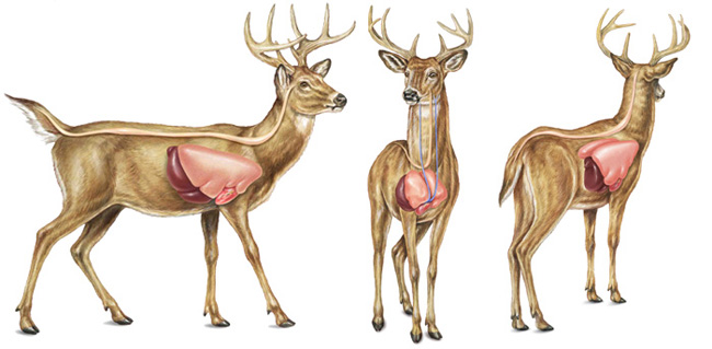
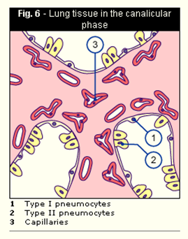
these are deer embryos from the fifth week, as you can see the heart is unseen, but is beating inside their bodies.
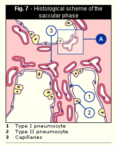
Zygote to the Bastula Stage


  • Zygote= a cell that is a result from fertilization.
  • Zygotes undergo mitotic cell divisions, or mitosis, to become an embryo.
  • The blastula= an early stage of embryonic development in animals. It is also called the blastosphere.
  • Cleavage= the division of cells in the early embryo. The zygotes go through rapid cell cycles with no significant growth and produces many cells the same size as the original zygote.
  • End of cleavage is known as midblastula transition
  • After cleavage has produced over 100 cells the embryo is called a blastula.

When Does the Heart Start Beating ?

Week 5
The embryo’s tiny heart begins to beat by day twenty-one.

Development of the Four Chambered Heart

As warm-blooded animals, deer's use a lot of energy and therefore need a great supply of oxygen for their activities. Thanks to the four-chambered heart, they are at an evolutionary advantage: they're able to roam, hunt and hide even in the cold of night, or the chill of winter.

Wednesday, June 12, 2013

Doctor's Notes 2
Now that we understand what passive transport is, let’s learn about active transport! 

  • Active transport always requires energy (ATP) 
  • Molecules move up the concentration gradient, in other words, molecules move from low to high concentration gradients. 
  • Has three forms; ion pumps (also known as Sodium-Potassium Pumps), endocytosis, and exocytosis. 
  • Sodium-Potassium Pumps- transport sodium and potassium ions up the concentration gradient.
  • Endocytosis- molecules/particles enter the cell membrane (ex. an amoeba goes through endocytosis when it eats a piece of algae)
  • Exocytosis- molecules/particles exit the cell and are released outside.


Sodium-Potassium Pumps


Endocytosis


Exocytosis

Doctor's Notes 1

In order for Yuki to grow and take in the proper nutrients, we need to understand how organic molecules enter and leave her cells. There are two types of movements for substances to move across the cell membrane, passive transport and active transport.

If membranes of an animal cell are selectively permeable and there are concentration gradients extant in some of the molecules but not in others, then there must be “transporters” that allow specific solutes to cross the membrane. 

  • Some substances are able to use passive transport to cross the cell membrane, this transport does not involve energy from the cell. 
  • Passive transport is high concentration to low concentration (substances move down their concentration gradient)
  • Three forms of passive transport; diffusion, osmosis, and facilitated diffusion.
  • Diffusion- molecules move from an area of higher concentration to an area of lower concentration until they reach equilibrium; a state of equality on both sides.
  • Osmosis- process in which free water molecules diffuse across the cell membrane. 
    • Water moves from areas of low solute concentration to areas of high solute concentration until the solute concentrations are in equilibrium. 
    • If the animals cell’s membrane has a lower solute concentration than outside of the membrane, then the outside solution is said to be hypertonic (water will mostly diffuse from the inside solution to the outside solution)
    • Hypertonic- if too much water leaves the cell, the cell will shrink or shrivel up, killing the cell.
    • Hypotonic areas are found when the solution of the membrane has a lower solute concentration than the inside of the cell. (water will mostly diffuse from the outside solution to the inside and if there is too much water inside the cell, the cell may swell and eventually lyse, or burst) 
  • Facilitated diffusion- molecules are assisted by special transport proteins. 
    • Uses ion channel proteins and carrier proteins, that are found in the cell membrane, and can transport substances into and out of the cell.
  • Ion channel proteins allow specific ions to pass through the membrane. 
    • Regulated by the cell and control the passage of substances that enter or leave the cell. 
    • Lastly, carrier proteins attach to specific molecules to move the molecules across the membrane.

Osmosis


Diffusion


Facilitated Diffusion
What Would Happen to the Lung Growth if Something Goes Wrong in the Cell?
If something went wrong in the cell, it may affect the lung growth. Some possible malfunctions may be...

  • Lung Tumors
  • Lung Cancer (note: Lung Tumors may result in Lung Cancer)
  • COPD (Emphysema and Chronic Bronchitis)
  • Asthma
What Organic Molecules are Needed to Help the Fetus Grow?
In order for the fetus to grow properly and for its lungs to function properly, the fetus must obtain the proper amount of organic molecules.
  • Lipids: structural component of cell membranes and a source of energy in the form of fats.
  • Proteins are involved in structural support of the body, body movement, and defense against harmful substances.
  • Carbohydrates create energy, allowing the fetus to grow and function normally.
    For more information on how organic molecules enter and leave the cell, visit refer back to the Doctor's Notes; or, click on this link that will bring you to the site: http://mountaincorepreserve.blogspot.com/search/label/Doctor%27s%20Notes

Growth of the Organ Over Gestation Time:
How does a single cell grow into a whole lung?
  1. Tissues are in a state of tension where all their cells “hang” on each other.
  2. They pull on a flexible matrix of fibrous proteins and sugars that surround and support every cell.
  3. Cells beneath the growing lungs release proteins that unravel and make small regions in the tensed matrix thinner.
  4. These spots stretch more than the rest of the matrix, causing a thinning.
  5. These areas are stretched more than neighboring parts of lung tissue.
  6. If cells feel that extra tension, they begin to grow.
  7. Cells will bud and branch outwardly, forming new sacs. But if the cells feel a decrease in tension, they stop the growth.
  8. Growing lungs need supplies of blood to function.
Dr.  Donald Ingber, a Judah Folkman Professor of Vascular Biology at the Medical School in Harvard University, has seen an extension of small, nearby blood vessels, also known as capillaries, growing in series with new air sacs. He and his team conducted an experiment to test whether or not adjusting the tension in developing mouse lungs would allow the lung tissue to bud outward; and, it did. For more information on his discoveries, click on the link below.
http://www.news.harvard.edu/gazette/2005/03.24/03-lung.html
Introduction to Lung Development:
This is a diagram of the different phases of lung development. It describes the approximate time period lungs develop in a fetus and at what stage the lungs are in at a certain time period. Also, below are some descriptions of the five different phases that occur during lung development. I will go into more detail about the development of lungs as we learn about this new topic of growth.
http://www.embryology.ch/anglais/rrespiratory/phasen01.html
Week 4-8: Embryonic Phase
Lung development in a fetus deer starts during the embryonic phase.
  • Takes place during the first four or five weeks of gestation.
  • Larynx, or voicebox, and the trachea, also known as the windpipe, begin to form.
  • Two small buds branch off of the trachea, which will later develop as lungs. 

Key:
1=Right main bronchus
5=Left main bronchus
http://www.embryology.ch/anglais/rrespiratory/phasen02.html ttp://www.livestrong.com/article/163935-fetal-lung-development-stages/

Week 5-17: Pseudoglandular Phase

  • Begin at around the 9th week of gestational period
  • The branching of the lungs increase and bud into more areas.
  • Each bud eventually becomes an independent respiratory unit, which is served by a bronchiole, and is surrounded by capillary vessels that bring blood to the lungs for oxygen.
This diagram represents what is occurring in the pseudoglandular phase, where the lungs resemble a gland. At the end of this phase, the precursors of the pneumocytes can be discerned in the respiratory sections as cubic epithelium.
ttp://www.livestrong.com/article/163935-fetal-lung-development-stages/
Week 16-26: Canalicular Phase
  • Occurs at approximately 25 weeks in of the gestational period
  • An air-blood barrier is created that allows oxygen to enter the blood in respiratory capillaries and carbon dioxide to flow out of respiratory capillaries and into the lungs.
  • Air transports tissues, where the tissues become discernible from other gas-exchanged tissues
    .
In this diagram, the type I pneumocytes differentiate out of the type II pneumocytes. The capillaries are also approaching the walls of the acini during this phase.
http://www.embryology.ch/anglais/rrespiratory/phasen04.html ttp://www.livestrong.com/article/163935-fetal-lung-development-stages/

Week 24-38: Saccular Phase

  • Occurs at about the 36th week of the gestational age.
  • Surfactant production begins in the lungs.
  • Surfactant is a soapy substance that helps keep the lung tissue from sticking to itself and prevents tearing during exhalation or lung compression.
  • Air sacs begin to fill
In the diagram, the capillaries multiply around the acini. They push close to the surface and form a common basal membrane with that of the epithelium.
http://www.embryology.ch/anglais/rrespiratory/phasen05.html ttp://www.livestrong.com/article/163935-fetal-lung-development-stages/

Week 36-Birth: Alveolar Phase

  • Occurs during the 36th week of the gestational age.
  • The lung development during this phase is characterized by the growth of bronchioles and air sacs, or alveoli.
  • The alveoli allows the gas-exchange tissues of the lungs to expand and make them capable of moving more air into the lungs as the fetus grows.
The diagram above shows that in the alveolar phase, the alveoli form from the terminal endings of the alveolar sacculi and with time, they increase in diameter.
ttp://www.livestrong.com/article/163935-fetal-lung-development-stages/
After Birth:
  • After birth, and during the first couple months
  • Lungs continue to grow and expand until it is at its maximum growth.
This diagram depicts what happens in the alveolar phase after birth. More alveoli form from the terminal endings of the alveolar sacculi and increase in diameter overtime. Furthermore, they are also delimited by secondary septa.
http://www.embryology.ch/anglais/rrespiratory/phasen06.html

Yuki's Family History


Yuki, Suki, and her parent's were all Brown Whitetail Deer's, but Yuki's father and his family were all Albino deers, meaning that they were white in color.

Tuesday, June 11, 2013

Exciting News !!!

Suki finally gave birth ! We are so excited, and so glad we know so much about taking care of a baby deer ! We were not able to film this event, but found a video that was the same. We believe it is a bit graffical, so be warned. It was a girl, so we decided to name her Yuki. Here are some pictures of her.

Heart Formation Video

here is a small overview of the formation of the heart, there is a short one that isnt as detailed as the longer one, but both are filled with good information 
Short One

<-------Long one

Monday, June 10, 2013

Heart Formation Week 7

http://php.med.unsw.edu.au/embryology/index.php?title=Heart_Outflow_Septation_Movie

Now at this point, after the outflow separation, the heart formation is near done

Heart Formation Week 6

http://php.med.unsw.edu.au/embryology/index.php?title=Heart_Atrial_Septation_Movie

A brief video on the Heart Atrial Septation

Heart Formation Week 5

http://php.med.unsw.edu.au/embryology/index.php?title=Heart_Realign_Movie

The above link is a brief video of the realignment of the heart during its formation

Heart Formation Week 4

http://php.med.unsw.edu.au/embryology/index.php?title=Heart_Looping_Movie

Click on the above link to see the looping of the heart

Formation of Heart Week 3

http://php.med.unsw.edu.au/embryology/index.php?title=Week_3_Development_Movie

Click on the link to see a brief video of the third week of the heart's formation.

Formation of the Heart

Advanced Heart Development Timeline.jpg

Here is a brief timeline of the formation of the heart

Tuesday, June 4, 2013

Fawn's Diet

We may run a preserve, but this is the first time we will be taking care of a baby deer ! We have no clue what it needs to eat, what conditions it can and cannot survive in, which is why I've set up an appointment for Dr. William Levington, a good friend of mine, who specializes in baby mammals, and has agreed to help us get ready for the fawn's arrival around June 10th. Just like we did for Scientist Elizabeth Vang, we will take notes on what Dr. Levington will tell us and post it here. Unfortunately, this will not be open to the public, so please look here for the notes.

Until Next Time~